English
Big Sale – Last Days for 20% Off!
542HOUR 48MINUTE 21SECOND
JAW CYST or TUMOR? What Does the

JAW CYST or TUMOR? What Does the "Shadow" on a Panoramic X-ray Mean?

8 December Mon, 2025

Dr. Ali Direnç Ulaşan – Oral and Maxillofacial Surgeon, Milim Dental Bursa

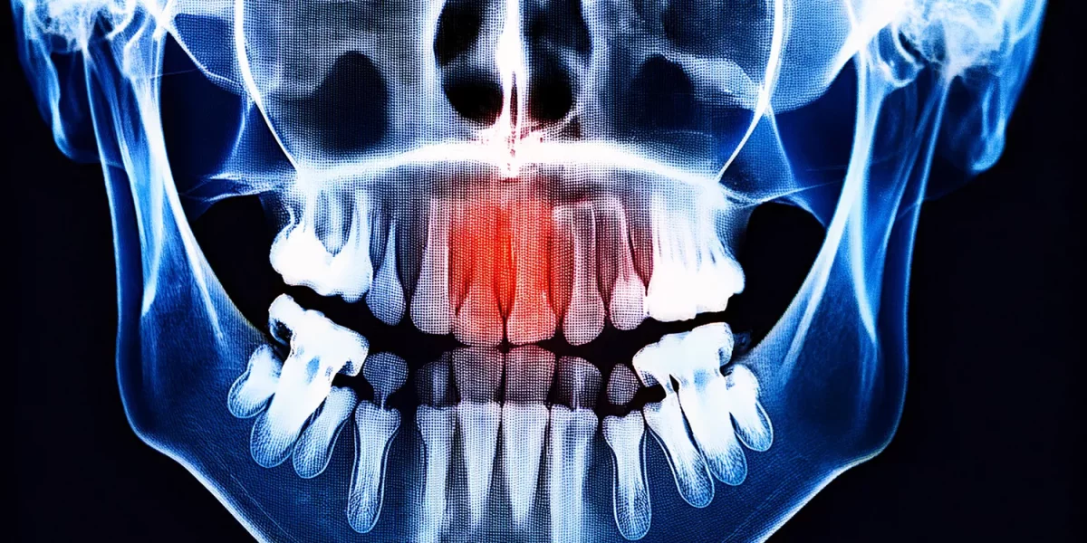
You go for a dental check-up, and a panoramic X-ray (a large jaw X-ray) is taken.

The report states:

Request a Free Treatment Plan from Our Expert Doctors

"A radiolucency (shadow) is observed in the right lower jaw region. Further examination is recommended for differentiation between a cyst/tumor."

And the only thought going through your mind is:

"Do I have a tumor? Am I cancerous?"

As a surgeon specializing in maxillofacial surgery in Bursa, I, Dr. Ali Direnç Ulaşan, and the Milim Dental team, see many patients who come to the clinic every day with exactly this worry.

In this article, I want to explain in a language that the public can understand that the "shadow" seen on a panoramic X-ray:

  1. Does not always mean a malignant tumor.
  2. Is often a cyst or a simple, manageable lesion.
  3. How the differentiation between a cyst and a tumor is made.
  4. In which situations one truly needs to be concerned.

1. What is the "Shadow" on a Panoramic X-ray?

First, let's clarify a basic point: The panoramic X-ray is the most common general screening X-ray used in dentistry, showing the entire mouth and jaws in a single image.

In this film:

  1. Areas seen as dense, nearly white: Usually structures like bone, fillings, metal, or implants.
  2. Areas seen as darker, nearly black: Areas where bone density is reduced or "empty" spaces, such as cystic/tumorous/normal cavities.

Therefore, the term mentioned in the report:

"Radiolucency" or "radiolucent area" simply means "a dark-appearing region on the X-ray."

This dark area can sometimes be:

  1. An ordinary anatomical space (e.g., the sinus cavity).
  2. A normal variation.
  3. A jaw cyst originating from a tooth.
  4. More rarely, a tumorous formation.

So, the phrase "there is a shadow" alone does not mean you have cancer. But it is a finding that should be taken seriously and evaluated by an oral and maxillofacial surgeon.

2. What is a Jaw Cyst? Is Every Cyst Dangerous?

A jaw cyst is a cavity within the jawbone or associated with tooth roots, filled with fluid or semi-fluid material, and surrounded by a thin membrane.

There are various types of jaw cysts, but their fundamental characteristics are:

  1. They are generally benign (non-cancerous).
  2. They grow slowly.
  3. They may remain asymptomatic for a long time.
  4. As they grow, they can push teeth, erode roots, and weaken the jawbone.

Common types of cysts (in lay terms):

  1. Apical (Root Tip) Cyst: Cysts seen at the root tip of infected teeth that require or have had root canal treatment.
  2. Cysts related to impacted teeth (Follicular/Dentigerous Cyst): Cysts developing around wisdom teeth or impacted canine teeth.
  3. Special cyst types like Keratocyst: Cysts with a higher tendency to recur, requiring closer follow-up.

The key takeaway here is: Not every jaw cyst is cancer; the vast majority are benign.

However, if left untreated, they can:

  1. Grow larger.
  2. Thin the jawbone, increasing the risk of fracture.
  3. Cause teeth to shift position and root resorption.

Therefore, it is not correct to leave them simply because they are "benign."

3. What is a Jaw Tumor? How Common are Malignant Ones?

Tumor is a general word we use for masses formed by uncontrolled cell proliferation. We classify them into two main categories:

  1. Benign Tumors:
  2. Progress more slowly.
  3. Usually do not infiltrate surrounding tissues; they are contained.
  4. Problems are often resolved when completely removed with appropriate surgery.
  5. Malignant Tumors / Cancers:
  6. Progress more aggressively.
  7. Tend to spread to surrounding tissues (invasion).
  8. Some can metastasize (lymph nodes, lungs, etc.).

A significant portion of tumors seen in the jaw area are benign.

Malignant jaw tumors and oral cancers are more commonly encountered in patients who:

  1. Have risk factors such as smoking, alcohol, poor oral hygiene, and chronic irritation.
  2. Often present with a complaint of a sore/mass on the tongue, inside the cheek, on the floor of the mouth, or on the palate.

Therefore, not every "shadow" seen on a panoramic X-ray during a routine check-up, when you have no complaints, means a "malignant tumor." However, the way to distinguish whether it is a cyst, a tumor, or a normal cavity is through detailed examination.

4. Does the "Shadow" Cause Symptoms? What Might the Signs Be?

Jaw cysts and some tumors can remain completely silent for a long time. This is why they are often not noticed until a routine panoramic X-ray is taken.

Sometimes, however, they can cause the following complaints:

  1. Feeling of fullness or swelling in the jaw.
  2. Pain during chewing.
  3. Tooth shifting, spacing, or tilting.
  4. Unexplained tooth looseness.
  5. Throbbing pain that doesn't subside despite inadequate root canal/filling treatments.
  6. In advanced cases, jawbone thinning or even fracture.

Much more rarely:

  1. Numbness in the lip or jaw area.
  2. A mass noticed inside or outside the mouth.
  3. A long-lasting, unhealed sore.

If you have such complaints, you must be evaluated by an oral and maxillofacial surgeon.

5. Jaw Cyst or Tumor? How is the Differentiation Made?

5.1. First Step: Clinical Examination

The first thing I do with patients who come to Milim Dental in Bursa is:

  1. Detailed intraoral and extraoral examination.
  2. Checking for swelling, tenderness, or color change.
  3. Performing tooth vitality tests.
  4. Checking for sensitivity with slight pressure on the gums, if necessary.

This step is crucial for combining the clinical findings with the shadow we see on the radiology.

5.2. Second Step: Radiological Examination (Panoramic + CBCT)

The panoramic X-ray gives us a general map. However, it is often insufficient on its own to determine the type of lesion. Therefore, especially in suspicious cases, we recommend getting a:

  1. CBCT (3D Dental Tomography).

Thanks to CBCT, we can see in 3D:

  1. The exact size of the lesion.
  2. Its location within the jawbone.
  3. Its relationship with important structures like the nerve canal, sinus cavity, and roots.
  4. Bone density.

Some types of cysts have typical appearances on radiographs (e.g., a well-defined, rounded space around an impacted wisdom tooth). Nevertheless, it is not always possible to say "it is 100% this just by looking at the radiological image."

5.3. Third Step: Biopsy and Pathology Examination

The actual critical differentiation lies here: "Is it a cyst, a tumor, benign, or malignant?"

The clear answer to this question is provided by the pathology laboratory. This means we surgically take a sample from the lesion (biopsy) or remove it completely; the tissue sample is then examined under a microscope.

Is a biopsy harmful?

The most common concern I hear from the public is: "Doctor, will it spread if you take a biopsy?"

No. A biopsy performed with the correct technique will not cause the disease to spread. On the contrary, by clarifying the diagnosis, it can prevent unnecessarily large surgeries.

In most cases of jaw cysts and benign tumors:

  1. The lesion is completely removed in one surgical session.
  2. The removed tissue is sent for pathology.
  3. Based on the result, it is evaluated whether an additional procedure is necessary.

6. How is a Jaw Cyst / Tumor Treated?

The treatment plan is determined based on:

  1. The type of the lesion.
  2. Its size.
  3. Its relationship with the jawbone.
  4. Its connection to the teeth.
  5. Your general health condition.

6.1. Jaw Cyst Treatment

The most common procedures we perform are:

  1. Enucleation (complete removal of the cyst): The cyst tissue and its surrounding membrane are cleaned out of the jawbone in one piece.
  2. Curettage: The thin walls in the area where the cyst was are cleaned by scraping (curettage).
  3. Marsupialization (window opening): In very large cysts, a part of the cyst is opened into the mouth and drained, and it is expected to shrink over time; it is then completely removed in a second surgery.

In some cases, the cavity created by the cyst may be supported with bone graft (bone powder). In the long term, once the area has recovered, implant planning can be done if necessary.

6.2. Benign Tumors

For benign jaw tumors, the goal is usually to:

  1. Completely remove the lesion within clear margins.
  2. If necessary, perform a resection along with a small amount of surrounding healthy bone.
  3. Subsequently, the jawbone form is restored with grafts and, if needed, implants/prostheses in the future.

6.3. Malignant Tumors and Oral Cancers

If the pathology result indicates a malignant tumor:

  1. A more comprehensive treatment plan is often developed, involving the collaborative work of oral and maxillofacial surgery, oncology, and radiation oncology.

Early detection of these cases is vitally important. Therefore, if you have:

  1. A sore in your mouth that lasts longer than 2 weeks,
  2. Unexplained bleeding,
  3. A hardened mass,
  4. Accompanying symptoms like pain and weight loss,

you should consult a specialist without delay.

7. What Should You Do When Told "There's a Shadow on the Panoramic X-ray"?

Don't panic, but don't ignore it either. A shadow does not always mean "tumor," but it requires detailed examination.

  1. Consult an oral and maxillofacial surgeon. Your general dentist may correctly refer you to a surgeon when they see a shadow on the panoramic X-ray. As Dr. Ali Direnç Ulaşan in Bursa, we routinely evaluate these cases at Milim Dental.
  2. Get a CBCT (3D tomography) if necessary. This provides much more detailed information about the size and location of the lesion.
  3. Do not postpone if your surgeon recommends a biopsy or surgical removal. The earlier the intervention, the smaller the lesion when resolved; the surgery is easier, and preserving the jaw structure is more feasible.

8. Dr. Ali Direnç Ulaşan's Approach for Jaw Cysts / Tumors in Bursa

At Milim Dental, we typically manage the process for patients presenting with suspected jaw cysts or tumors as follows:

  1. Detailed History and Examination:
  2. How long you've been aware of it.
  3. Your complaints (pain, swelling, numbness).
  4. Past root canal, filling, or implant procedures.
  5. Your systemic diseases (diabetes, osteoporosis, etc.).
  6. Panoramic Film + CBCT Examination:
  7. We create a 3D map of the lesion.
  8. Clearly Explaining the Treatment Plan:
  9. Is the likelihood of a cyst or a tumor higher?
  10. What type of surgery will be required?
  11. Is local anesthesia, sedation, or general anesthesia appropriate?
  12. How will your teeth and implant needs be planned afterward?
  13. Surgical Procedure and Pathology Follow-up:
  14. Removal of the lesion.
  15. Control and long-term planning after the pathology result is received.
  16. Follow-up and Protective Approach:
  17. Some cyst types may have a tendency to recur. Therefore, we recommend:
  18. Panoramic X-ray or CBCT control at regular intervals.
  19. Clinical examination, if necessary.

9. Frequently Asked Questions (FAQ)

1) If a shadow appeared on the panoramic X-ray, is it definitely a cyst?

No. The shadow could be a cyst, tumor, normal cavity, anatomical variation, or an inflammatory focus. Clinical examination, CBCT, and biopsy (if necessary) are essential for clear differentiation.

2) If I have a cyst, do I have to have surgery immediately?

It depends on the type, size, and effect on surrounding tissues. While small, incidentally found lesions can be monitored, surgery is necessary for cysts that pose a risk of bone fracture, push teeth, or approach structures like nerves and sinuses. You will make this decision with your oral and maxillofacial surgeon.

3) I am afraid of having a biopsy; will it spread?

A biopsy taken with the correct surgical technique will not cause the disease to spread. On the contrary, it ensures an accurate diagnosis, preventing unnecessary or inadequate treatments.

4) Can a jaw cyst recur?

Some cyst types (e.g., keratocyst) have a higher risk of recurrence than others. Therefore, we recommend regular check-ups based on the cyst type in your pathology report. Regular follow-up allows us to catch any potential recurrence early.

5) What if it turns out to be malignant?

If the pathology result shows a malignant tumor, we will refer you to a process managed collaboratively by the oral and maxillofacial surgery, oncology, and, if necessary, plastic surgery teams. Early diagnosis significantly increases the chance of treatment and quality of life. Therefore, early consultation and not avoiding a biopsy are very important.

10. Conclusion: The "Shadow" on the Panoramic X-ray is a Warning, Not a Verdict

In summary:

  1. The "shadow" seen on a panoramic X-ray does not mean "you have cancer."
  2. It is often a benign jaw cyst or a manageable lesion.
  3. However, you should not dismiss it as "it's probably nothing." Because it can grow, weaken your jawbone, and lead to tooth loss.

For a definitive diagnosis, the safest route is: oral surgeon examination + 3D tomography + biopsy (if necessary).

If you have:

  1. Learned that a "shadow" was seen on your panoramic X-ray,
  2. Feel an unexplained swelling or fullness in your jaw,
  3. Been described a suspicious area next to an impacted tooth or an old root-canal-treated tooth,

As Dr. Ali Direnç Ulaşan and the Milim Dental team, specializing in oral and maxillofacial surgery in Bursa, we are here to create the most accurate diagnosis and treatment plan for you together.

This article was prepared not to scare you, but to calm you with information and encourage informed action. The best decision is always made through timely professional examination.
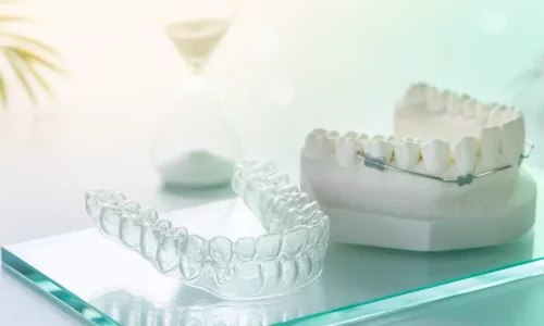

A Milim
Makes a Big Difference

Milim Dental Hospital is the first dental centre to receive the "Health Tourism Authorisation Certificate" issued by the Ministry of Health of the Republic of Turkey.
All Articles
Shine Like a Star: Hollywood Smile with E-Max Veneers at Milim Dental in Bursa
east

A perfect smile isn’t just about white teeth — it’s about harmony, confidence, and individuality. At Milim Dental, located in the serene city of Bursa, Turkey, we create dazzling, natural-looking transformations with E-Max veneers, bringing you one step closer to the legendary Hollywood Smile.

Hollywood Smile in Turkey: A Glamorous Transformation Awaits You in Bursa
east

When it comes to combining a stunning smile makeover with a luxurious and affordable vacation, Dent in Turkey has become a global phenomenon — and for good reason. With its high standards in dental care and cost-effective treatment options, Turkey is now the go-to destination for patients from Europe, the Middle East, and beyond. Among the standout cities in this dental revolution, Bursa shines as a unique blend of tradition, innovation, and wellness.

The All-on-X Procedure: Step-by-Step Guide
east

The All-on-X procedure involves several key steps, including a detailed consultation, implant placement, temporary prosthesis, healing and integration, followed by the permanent prosthesis. This comprehensive approach ensures a smooth recovery and successful outcome for full-arch restoration.

Why Patients
Choose Milim?

Milim Dental Hospital isn't just a clinic—it's where confident smiles begin. With a team of world-class specialists, advanced technology, and a patient-first approach, we turn dental care into a premium experience.
We prioritize hygiene, comfort, and tailor-made treatments designed just for you. Don’t just take our word for it—explore real stories from real patients.
Your perfect smile starts here. Join the Milim experience.

View All Experiences
play_arrow
Dt. İsmail Özkısaoğlu Dentist
Get a Free Consultation
play_arrow
Dr. Dt. Ali Direnç Ulaşan Oral, Dental, Maxillofacial Surgeon
Get a Free Consultation
play_arrow
Dr. Dt. Ali Direnç Ulaşan Oral, Dental, Maxillofacial Surgeon
Get a Free Consultation

Milim Dental Hospital provides comprehensive dental services in a spacious 1,000 m² facility, supported by a wide team of dental professionals including specialists in Oral and Maxillofacial Surgery, Prosthodontics, Orthodontics, Pediatric Dentistry, and Periodontology.

Our clinic is authorized by the Turkish Ministry of Health for International Health Tourism. This website is intended only for informing patients from abroad. It does not contain advertisements and complies with legal privacy regulations.
Milim Dental Hospital © 2025 - All rights reserved.

Have a Question on Your Mind?

English
Big Sale – Last Days for 20% Off!